清醒小动物功能超声成像系统
仪器介绍
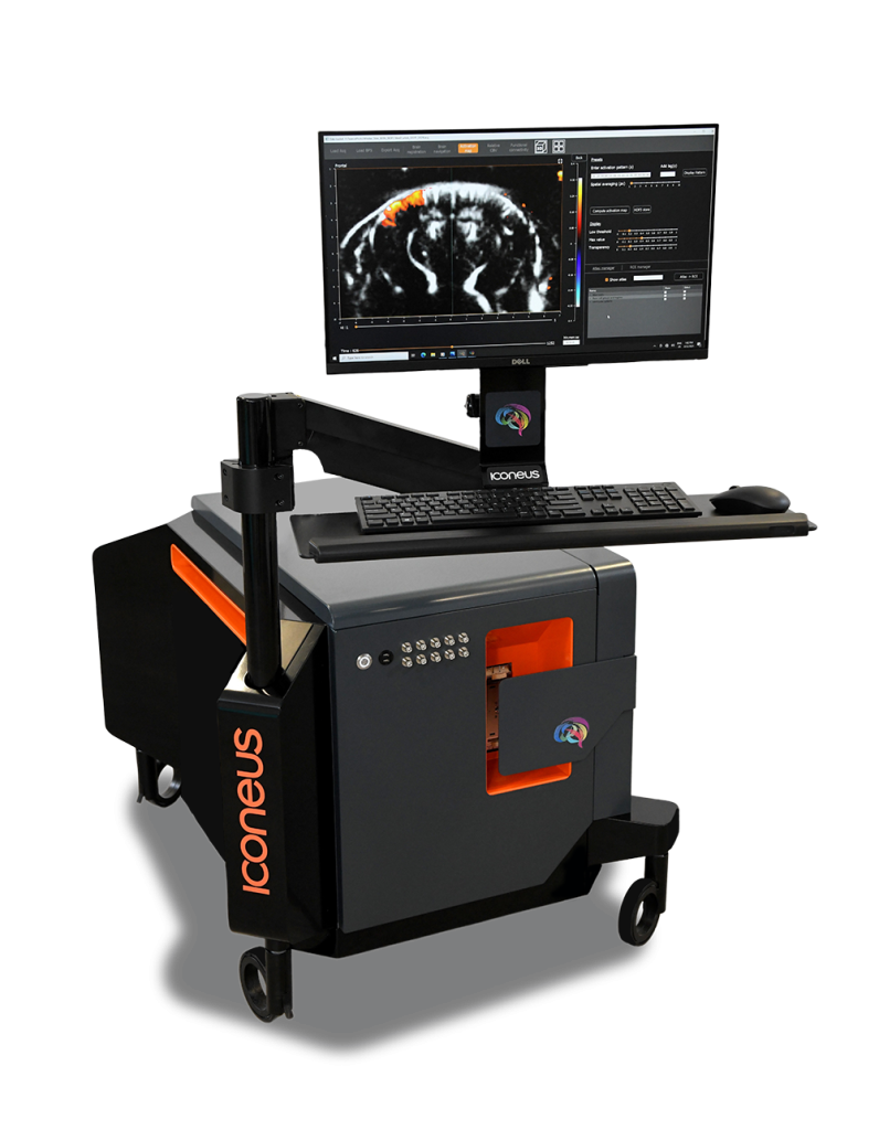
Iconeus One是一款功能性超声(fUS)脑成像系统,采用"多平面"超声波技术,可实现10 ms的高时间分辨率和100 μm的高空间分辨率。配备的超声定位显微镜(ULM)功能可实现5 μm的超高分辨率,并可量化mm/s级的血流速度。此外,系统配备了64、128和256通道的低噪音超轻探头,以及用于小动物头部固定扫描定位的电动四轴移动装置,可实现自动化、多角度扫描成像。软件内置了Allen小鼠脑参考图谱,能够自动识别并标记脑区,使数据分析更加高效和标准化。Iconeus One适用于从啮齿类到非人灵长类等各种动物模型,能够在头部固定或自由活动状态下进行实时、大视场、高灵敏度的2D/3D脑功能成像,是神经科学研究的有力工具。
参数
| Main unit | ICONEUS-ONE 256 channel |
| Probes | a) IcoPrime-4D MultiArray 15/256: for Head-fixed mice, multi-slice b) IcoPrime 15/128: for both head-fixed and mobile animals, single slice c) IcoPrime-Lite 15/128: for Mobile rats, single slice d) IcoPrime-Mini 15/64: for Mobile mice, single slice |
| Scanning platform | Four-axis motorized scanning platform for anesthetized or head fixed animals |
| Super resolution module | With specific imaging sequence for data acquisition and calculation module for post processing, Ultrasound Localization Microscopy (ULM) uses fUS to track injected microbubbles within blood vessels. This allows images of the micro-vasculature to be generated, with resolution as low as 5 µm. |
| Spatial resolution | Default mode: 100 × 100 × 400 μm at 15 MHz, with depth of ~1.5 cm ULM mode: Imaging micro-vasculature with resolution as low as 5 µm. |
| Software | a) IcoScan:Data live visualization and high-sensitivity single/multi-slice acquisition, 2D/3D angiography, B-mode imaging option. b) IcoStudio: Neuro-navigation function with automatic atlas registration (Allen Mouse Brain Reference Atlas integrated) and streamlined probe positioning; 2D/3D data visualization and analysis for functional ultrasound including activation map, relative CBV changes and functional connectivity. c) IcoLab: Batch processing for functional ultrasound analysis, including seed mapping and connectivity matrices, with report generation; Configurable workflows for ULM map computation for advanced vascular super-resolution studies. |