Imaging
The Imaging Core at BioCRF is a leading facility that offers access to various forefront imaging equipment and cutting-edge analysis tools for researchers. The Core comprehensively integrates diversified optical imaging technologies, including wide-field imaging, total internal reflection fluorescence imaging, confocal microscopy, spinning disk confocal, high-content screener, light sheet, two-photon, slide scanner, atomic force microscopy, live cell imaging, and super-resolution imaging. Additionally, the Core provides professional image processing workstations and image analysis software, empowering researchers to efficiently process and interpret their imaging data. The introduction of MINFLUX technology represents a significant breakthrough in the field of microscopy with its remarkable 2-nanometer super-resolution capability. This innovative technology opens up new opportunities for researchers to explore the intricate and previously inaccessible realms of the nanoscale world, providing unprecedented detail and precision in visualizing subcellular structures.
Staff in charge: Dr. Jianwei LIU and Ms. Siyi LI

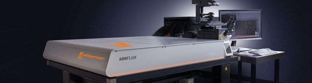
MINFLUX Super-Resolution Microscope
Model:
MINFLUX
Location:
123A, 1F, E4
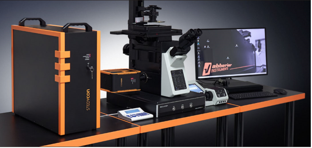
STED Super-Resolution Microscope
Model:
STEDYCON
Location:
123A, 1F, E4
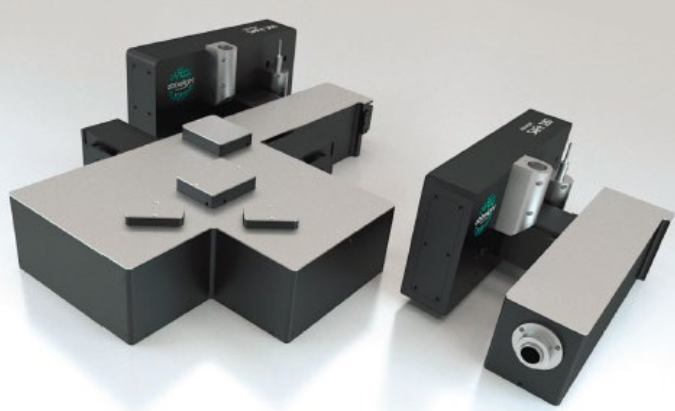
Single Molecule Localization Super Resolution System
Model:
SAFe 360
Location:
123A, 1F, E4
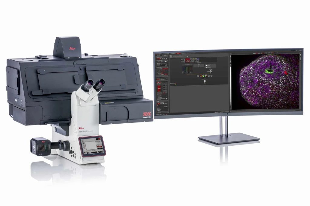
High-resolution live cell imaging system
Model:
DMi8 automated with Thunder
Location:
123C, 1F, E4
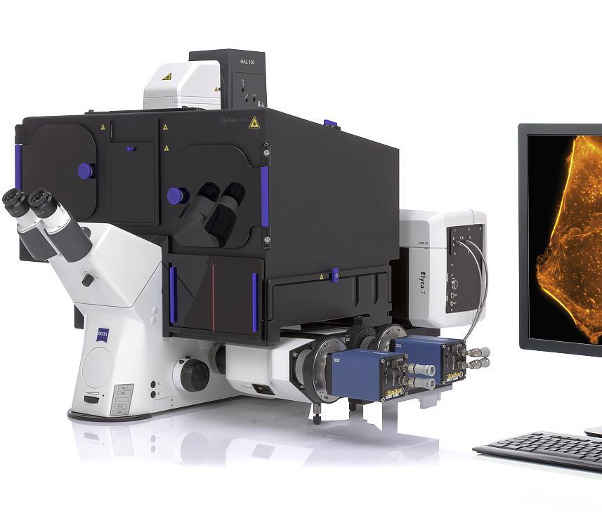
Super-resolution Microscope
Model:
Elyra 7
Location:
123B, 1F, E4
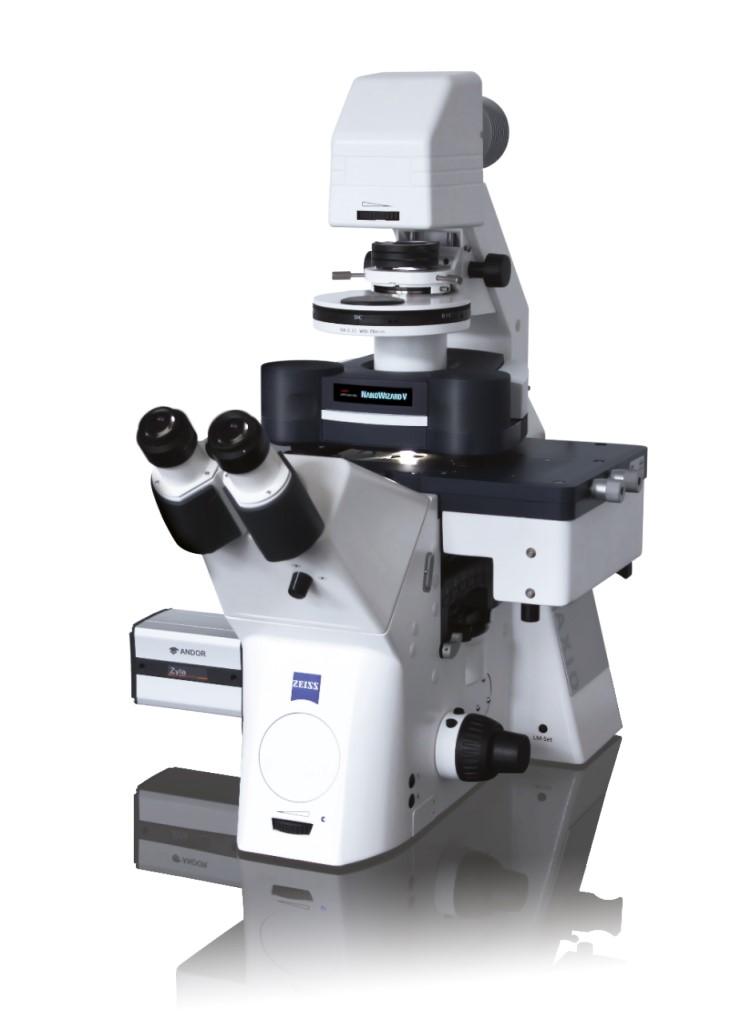
Atomic Force Confocal Microscope
Model:
NanoWizard V
Location:
123D, 1F, E4
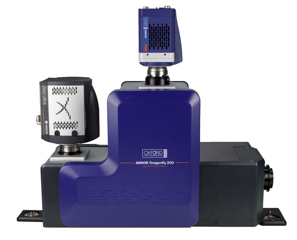
Spinning Disk High-speed Confocal Imaging System
Model:
Dragonfly 202
Location:
123B, 1F, E4
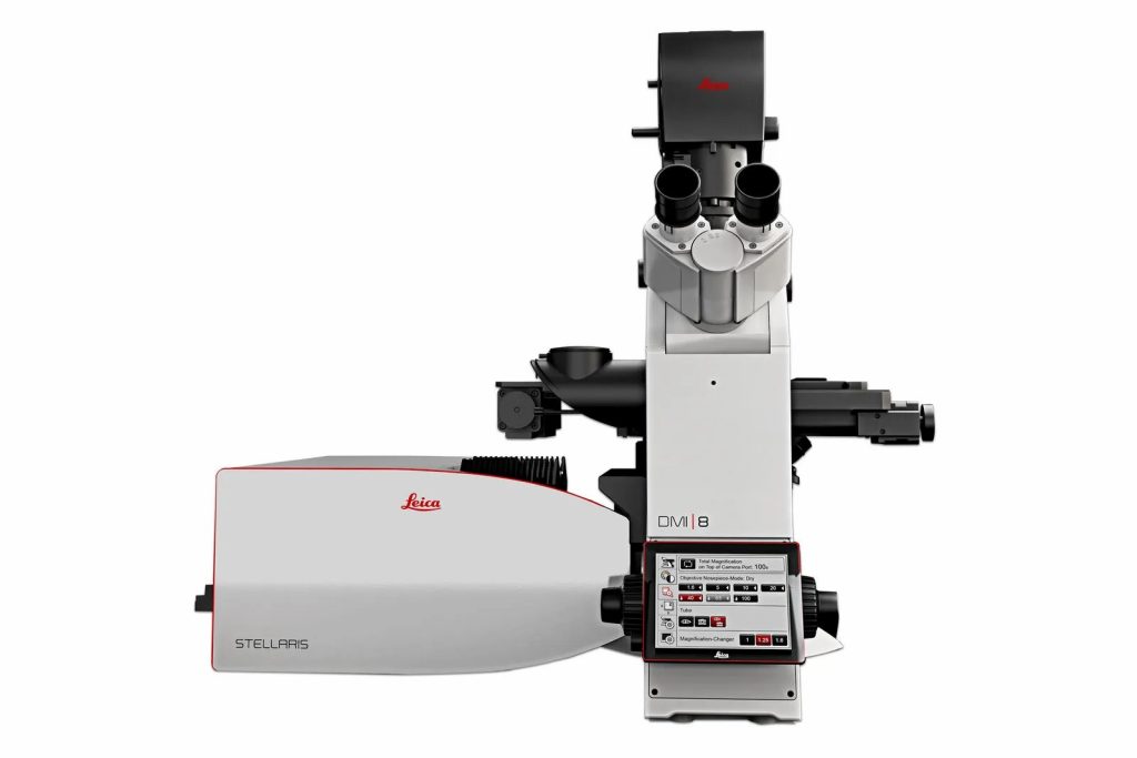
Laser Confocal Microscope
Model:
STELLARIS 5
Location:
123B, 1F, E4
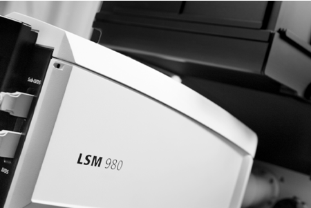
LSM 980 Confocal Microscope
Model:
LSM 980
Location:
123B, 1F, E4
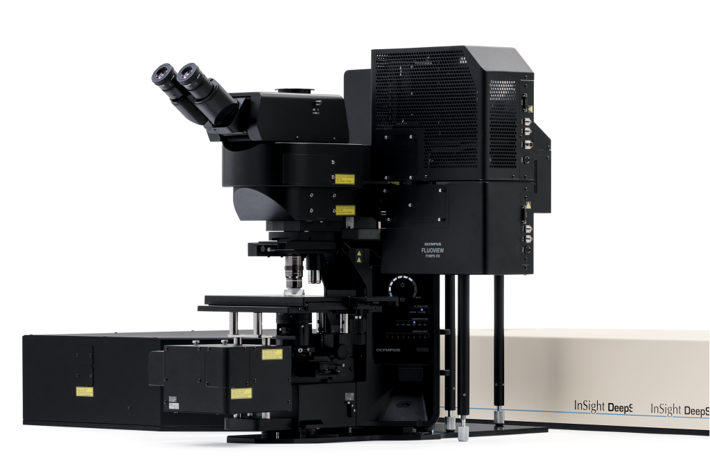
Two-photon Microscopy lmaging System
Model:
FVMPE-RS
Location:
128C, 1F, E4