Imaging
The Imaging Core at BioCRF is a leading facility that offers access to various forefront imaging equipment and cutting-edge analysis tools for researchers. The Core comprehensively integrates diversified optical imaging technologies, including wide-field imaging, total internal reflection fluorescence imaging, confocal microscopy, spinning disk confocal, high-content screener, light sheet, two-photon, slide scanner, atomic force microscopy, live cell imaging, and super-resolution imaging. Additionally, the Core provides professional image processing workstations and image analysis software, empowering researchers to efficiently process and interpret their imaging data. The introduction of MINFLUX technology represents a significant breakthrough in the field of microscopy with its remarkable 2-nanometer super-resolution capability. This innovative technology opens up new opportunities for researchers to explore the intricate and previously inaccessible realms of the nanoscale world, providing unprecedented detail and precision in visualizing subcellular structures.
Staff in charge: Dr. Jianwei LIU and Ms. Siyi LI


Free-Moving Triple Calcium Imaging Two-Photon Fluorescence Microscope
Model:
SUPERNOVA 600
Location:
128B, 1F, E4
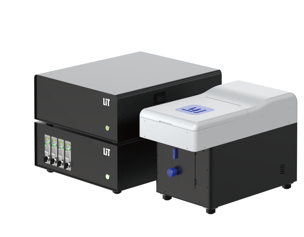
Large Sample Light-sheet Microscope
Model:
LiTone XL
Location:
123G, 1F, E4
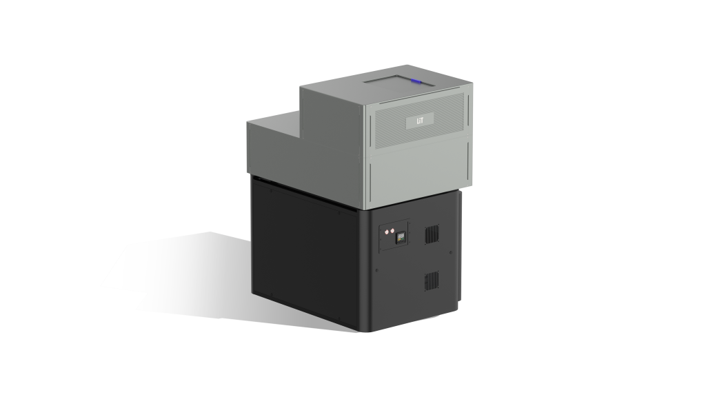
Live Sample Light-sheet Microscope
Model:
LiTone LBS2
Location:
123H, 1F, E4
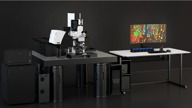
Full-spectrum High-resolution Fluorescence Lifetime Laser Confocal Microscope
Model:
STELLARIS 8
Location:
123E, 1F, E4
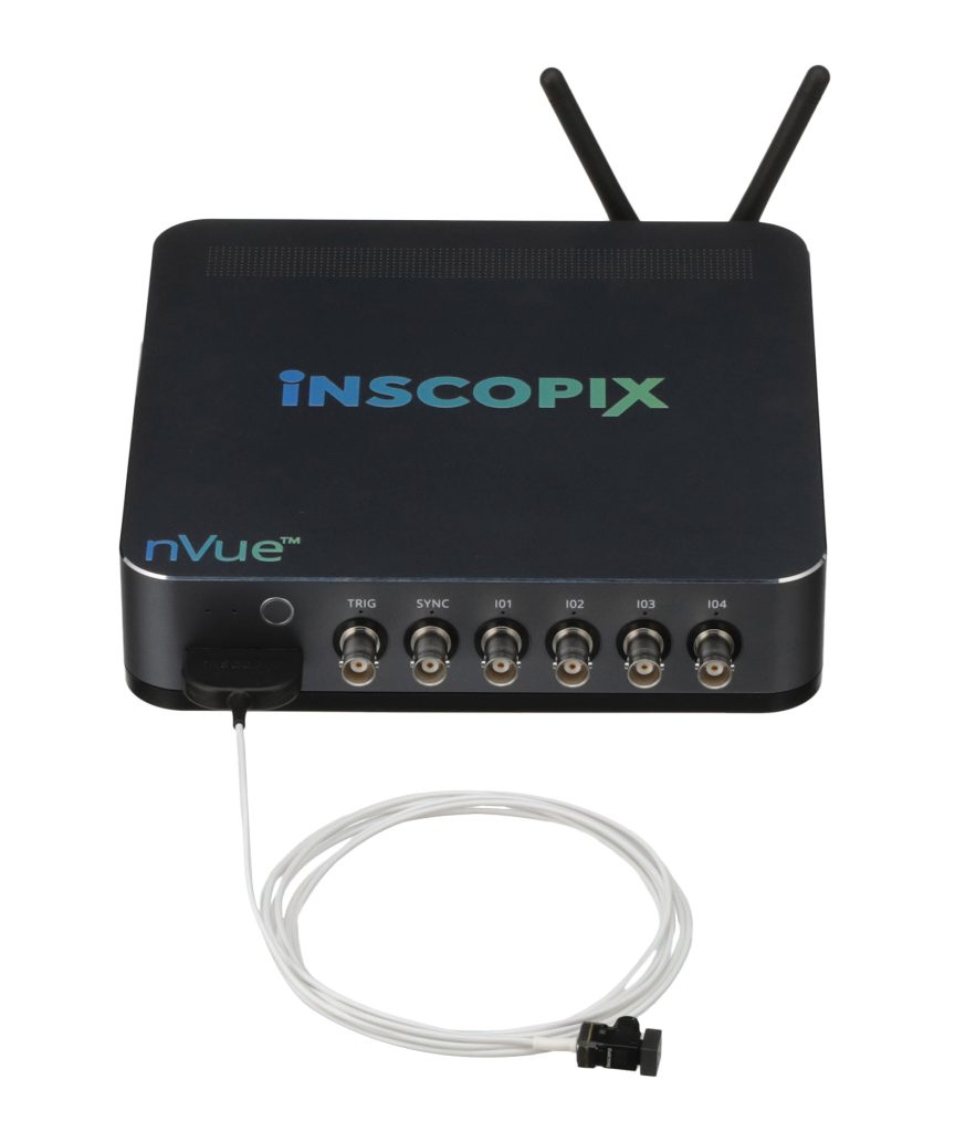
Dual-Color Head-Mounted Mini-Microscope Imaging System
Model:
nVue Lscape
Location:
128C, 1F, E4
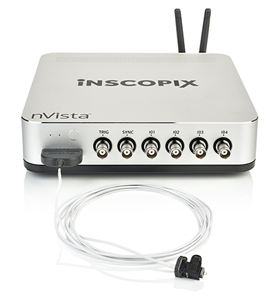
Monochromatic Head-Mounted Mini-Microscope Imaging System
Model:
nVista Lite
Location:
128C, 1F, E4
Laser Microdissection and Single-Cell Picking System
Model:
MMi CellCut Plus & CellEctor
Location:
128A, 1F, E4
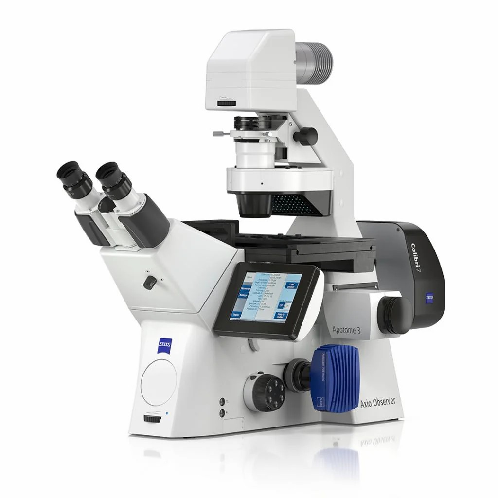
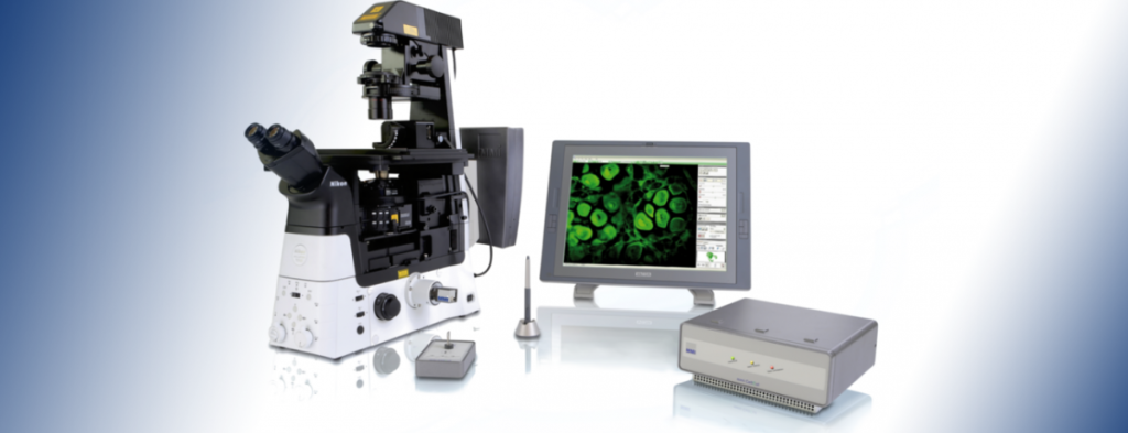
High-Resolution Inverted Fluorescence Microscope
Model:
Axio Observer 7 with Apotome3
Location:
123C, 1F, E4
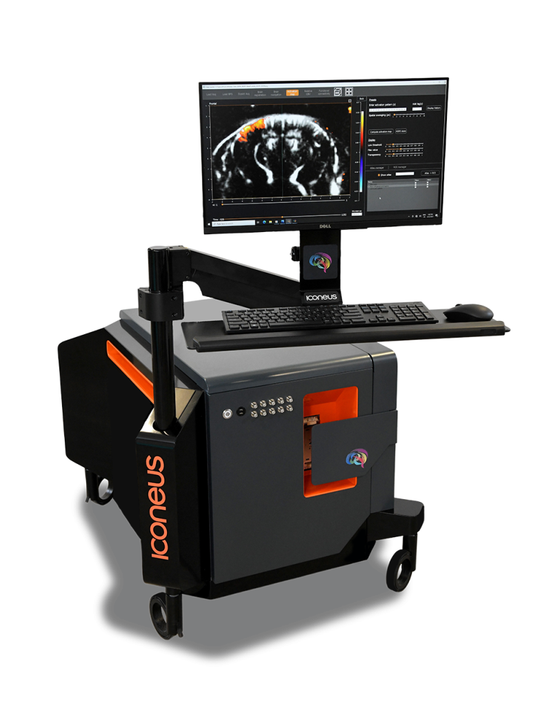
Small Animal Functional Ultrasound Imaging System
Model:
ICONEUS ONE
Location:
128B, 1F, E4
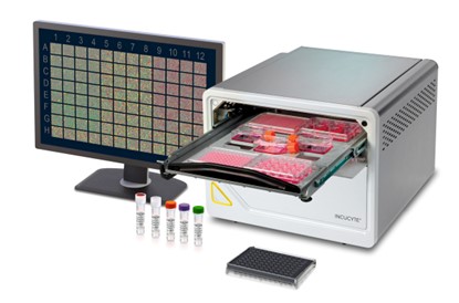
Live-cell Analysis System
Model:
Incucyte SX5
Location:
126, 1F, E4